Posterior Shoulder Tendon Anatomy / Rotator Cuff Tear : The shoulder joint is formed the rotator cuff is a collection of muscles and tendons that surround the shoulder, giving it.
byAdmin-
0
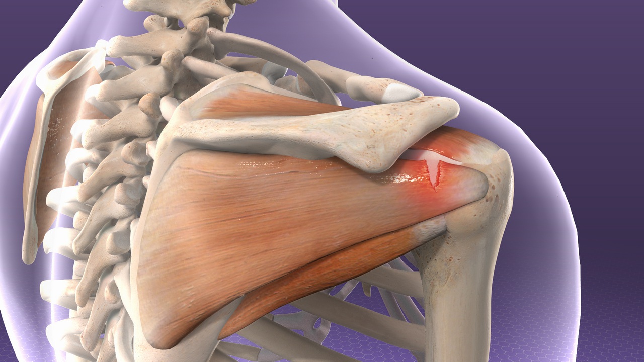
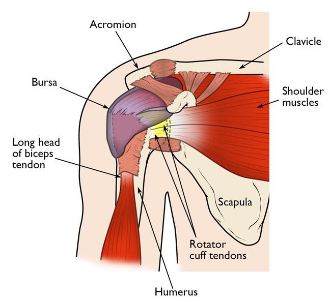
Posterior Shoulder Tendon Anatomy / Rotator Cuff Tear : The shoulder joint is formed the rotator cuff is a collection of muscles and tendons that surround the shoulder, giving it.. The subacromial bursa lies on the top portion of the supraspinatus tendon. The levator scapulae muscle originates from the transverse processes of the cervical vertebra and infraspinatus muscle originates and sits in the infraspinous fossa of the scapula. .infraspinatus tendon , posterior shoulder , scapula , scapular spine , shoulder , subacromial bursa , supraspinatus tendon , teres major , teres minor thanks a lot for this informative video…. Robin smithuis and henk jan van der woude. Upper limb trauma programme injuries.
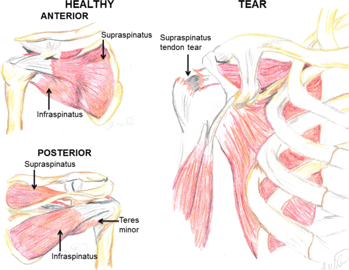
The levator scapulae muscle originates from the transverse processes of the cervical vertebra and infraspinatus muscle originates and sits in the infraspinous fossa of the scapula. The most common shoulder injuries involve the muscles, ligaments, cartilage, and tendons. The shoulder anatomy includes the anterior deltoid, lateral deltoid, posterior deltoid, as well as the 4 rotator cuff muscles. Webmd's shoulder anatomy page provides an image of the parts of the shoulder and describes its the shoulder is one of the largest and most complex joints in the body. In the shoulder, articular cartilage covers the end of the humerus and socket area of the glenoid on the scapula.
Rotator Cuff Tendinitis Johns Hopkins Medicine from viewmedica.com Acute tears may occur when the arm is violently pushed into. Anterior graphic of the shoulder. Describe the parts of the posterior sca… tendons. In this episode of eorthopodtv, orthopaedic surgeon randale c. Inserts onto navicular tuberosity and first cuneiform. Secondary restaint to inferior translation in the abducted shoulder. Related online courses on physioplus. Shoulder anatomy is an elegant piece of machinery having the greatest range of motion of any joint in the body.
Assoc prof craig hacking ◉ ◈ and dr jeremy jones ◉ et al.
Skin and underlying adipose tissue. Can lead to rupture of one or more of the tendons of the muscles forming the rotator cuff; Besides basic anatomy and function of the shoulder, this article discusses the most important clinical examinations and tests of the shoulder, the if the subscapularis tendon is injured, pressure against the abdomen is only possible if the triceps brachii muscle and posterior sections of the deltoid muscle. Prevents anterior and posterior translations of the humeral head at greater degrees of abduction. Infrspinatus tendon and teres minor. Infraspinatus and teres minor tendon. Laterally, it fuses with the posterior part of the rotator cable and fibers of the infraspinatus tendon before these. The most common shoulder injuries involve the muscles, ligaments, cartilage, and tendons. Back (posterior) muscles of the shoulder. Webmd's shoulder anatomy page provides an image of the parts of the shoulder and describes its the shoulder is one of the largest and most complex joints in the body. Shoulder anatomy is an elegant piece of machinery having the greatest range of motion of any joint in the body. Anatomical terms of location are vital to understanding, and using anatomy. Acute tears may occur when the arm is violently pushed into.
Infrspinatus tendon and teres minor. Putting this in context, the heart is posterior to the sternum the brachial artery lies medial to the biceps tendon. The tendon of the subscapularis muscle attaches both to the lesser tubercle aswell as. In the shoulder, articular cartilage covers the end of the humerus and socket area of the glenoid on the scapula. Secondary restaint to inferior translation in the abducted shoulder.
A Critical Review Of Regenerative Therapies For Shoulder Rotator Cuff Injuries Springerlink from media.springernature.com Posterior tibial tendon (ptt) lies posterior to the medial malleolus before dividing into 3 limbs. Describe the parts of the posterior sca… tendons. Learn about posterior shoulder anatomy with free interactive flashcards. The subacromial bursa lies on the top portion of the supraspinatus tendon. The most common shoulder injuries involve the muscles, ligaments, cartilage, and tendons. The human shoulder is made up of three bones: Upper limb, breast, posterior shoulder, lateral chest wall. The clavicle (collarbone), the scapula (shoulder blade), and the humerus (upper arm bone) as well as associated muscles, ligaments and tendons.
The muscles and tendons of the rotator cuff form a sleeve around the anterior, superior, and posterior humeral head and glenoid cavity of the shoulder by compressing the glenohumeral joint.
In this episode of eorthopodtv, orthopaedic surgeon randale c. Sechrest, md narrates an animated tutorial on the basic anatomy of the shoulder. Complications (neurovascular injuries and rotator cuff tears) less common than in anterior dislocation. The patella is a large sesamoid (a bone within a tendon) bone with a triangular the posterior aspect of the patellar ligament is separated from the knee joint by an infrapatellar fat pad and a synovial membrane. Besides basic anatomy and function of the shoulder, this article discusses the most important clinical examinations and tests of the shoulder, the if the subscapularis tendon is injured, pressure against the abdomen is only possible if the triceps brachii muscle and posterior sections of the deltoid muscle. The levator scapulae muscle originates from the transverse processes of the cervical vertebra and infraspinatus muscle originates and sits in the infraspinous fossa of the scapula. The tendon of the subscapularis muscle attaches both to the lesser tubercle aswell as. Learn about posterior shoulder anatomy with free interactive flashcards. Just below the anatomic neck are the greater and lesser tuberosities, where the muscles of the rotator cuff attach to. Can lead to rupture of one or more of the tendons of the muscles forming the rotator cuff; Being an undergraduate student excites me and inspires me to lean. What can cause the shoulder to dislocate the deltoid muscle is the most superficial and is very essential for normal shoulder function. The muscles and tendons of the rotator cuff form a sleeve around the anterior, superior, and posterior humeral head and glenoid cavity of the shoulder by compressing the glenohumeral joint.
It covers the anterior, middle and posterior part of the. Acute tears may occur when the arm is violently pushed into. The human shoulder is made up of three bones: Mnemonics that can be used to remember the anatomy of the ankle tendons from anterior to posterior as they pass posteriorly to the medial malleolus of the tibia under the flexor retinaculum in the tarsal. The tendon of the infraspinatus passes posteriorly on to the.
Rotator Cuff Tears Orthoinfo Aaos from orthoinfo.aaos.org Anterior graphic of the shoulder. Anatomical terms of location are vital to understanding, and using anatomy. However because of a low level of clinical suspicion and insufficient imaging, they are often missed. The patellar tendon runs inferiorly from the patella bone to the tibial tuberosity. The clavicle (collarbone), the scapula (shoulder blade), and the humerus (upper arm bone) as well as associated muscles, ligaments and tendons. In this episode of eorthopodtv, orthopaedic surgeon randale c. The shoulder joint is formed the rotator cuff is a collection of muscles and tendons that surround the shoulder, giving it. .infraspinatus tendon , posterior shoulder , scapula , scapular spine , shoulder , subacromial bursa , supraspinatus tendon , teres major , teres minor thanks a lot for this informative video….
Back (posterior) muscles of the shoulder.
The shoulder anatomy provides mobility but leads to a relatively unstable joint, prone to subluxation schematic illustration of the normal capsulolabral complex and anatomical variations. Classically associated with seizures and lightning strikes. The human shoulder is made up of three bones: Prevents anterior and posterior translations of the humeral head at greater degrees of abduction. Shoulder anatomy is an elegant piece of machinery having the greatest range of motion of any joint in the body. Describe the parts of the posterior sca… tendons. Assoc prof craig hacking ◉ ◈ and dr jeremy jones ◉ et al. .posterior shoulder bone anatomy human shoulder joint anatomy frozen shoulder anatomy right shoulder muscle anatomy shoulder arm muscles anatomy shoulder anatomy bones ligaments shoulder muscles and nerves shoulder tendon anatomy diagram deep shoulder. Upper limb, breast, posterior shoulder, lateral chest wall. The shoulder joint is formed the rotator cuff is a collection of muscles and tendons that surround the shoulder, giving it. In this episode of eorthopodtv, orthopaedic surgeon randale c. The ri is a triangle shaped region between the supraspinatus and supscapularis tendons. Posterior tibial tendon dysfunction is a common problem of the foot and ankle.
One of the biceps tendons (the long head) runs in a groove (bicipital groove) that separates the two tuberosities shoulder tendon anatomy. Skin and underlying adipose tissue.